Home

söz vermek yazılım iz mouse embryo atlas en yüce meleklerden biri çocuklar renk

Embryology of the mouse

Three-dimensional microCT imaging of mouse development from early  post-implantation to early postnatal stages - ScienceDirect
Three-dimensional microCT imaging of mouse development from early post-implantation to early postnatal stages - ScienceDirect

Schematic of the algorithm for identifying whole organ volume... | Download  Scientific Diagram
Schematic of the algorithm for identifying whole organ volume... | Download Scientific Diagram

Three-Dimensional Analysis of Vascular Development in the Mouse Embryo |  PLOS ONE
Three-Dimensional Analysis of Vascular Development in the Mouse Embryo | PLOS ONE

eMouseAtlas: An atlas-based resource for understanding mammalian  embryogenesis - ScienceDirect
eMouseAtlas: An atlas-based resource for understanding mammalian embryogenesis - ScienceDirect

eMouseAtlas: An atlas-based resource for understanding mammalian  embryogenesis - ScienceDirect
eMouseAtlas: An atlas-based resource for understanding mammalian embryogenesis - ScienceDirect

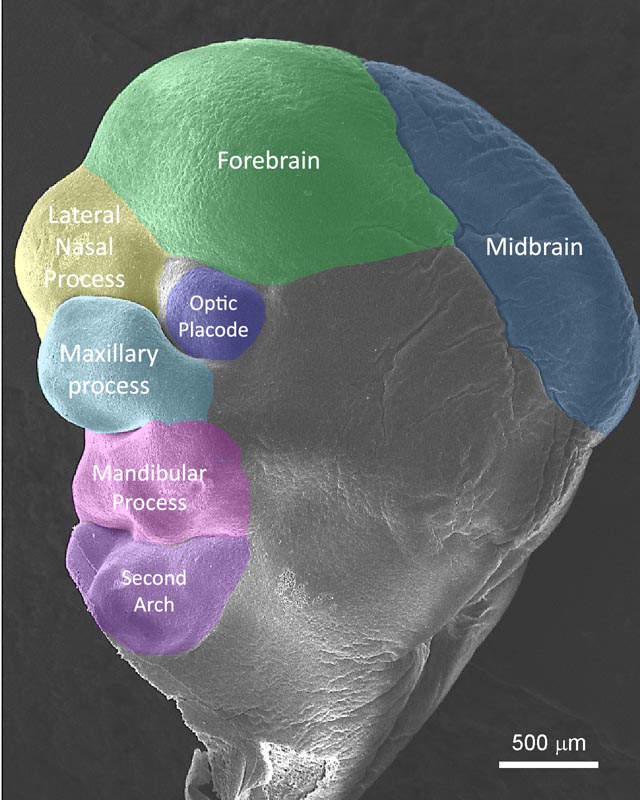
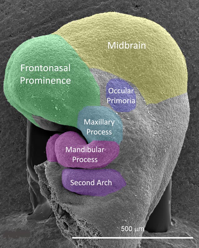
Mouse Anatomy | FaceBase

Mouse RNA Atlas
Mouse RNA Atlas

EMAP Home
EMAP Home

High-resolution magnetic resonance histology of the embryonic and neonatal  mouse: A 4D atlas and morphologic database | PNAS
High-resolution magnetic resonance histology of the embryonic and neonatal mouse: A 4D atlas and morphologic database | PNAS

High-resolution magnetic resonance histology of the embryonic and neonatal  mouse: A 4D atlas and morphologic database | PNAS
High-resolution magnetic resonance histology of the embryonic and neonatal mouse: A 4D atlas and morphologic database | PNAS

LAMA: Automated image analysis for developmental phenotyping of mouse  embryos | bioRxiv
LAMA: Automated image analysis for developmental phenotyping of mouse embryos | bioRxiv

PDF] 4D atlas of the mouse embryo for precise morphological staging |  Semantic Scholar
PDF] 4D atlas of the mouse embryo for precise morphological staging | Semantic Scholar

Histology Atlas of the Developing Prenatal and Postnatal Mouse Central  Nervous System, with Emphasis on Prenatal Days E7.5 to E18.5 - Vivian S.  Chen, James P. Morrison, Myra F. Southwell, Julie F.
Histology Atlas of the Developing Prenatal and Postnatal Mouse Central Nervous System, with Emphasis on Prenatal Days E7.5 to E18.5 - Vivian S. Chen, James P. Morrison, Myra F. Southwell, Julie F.
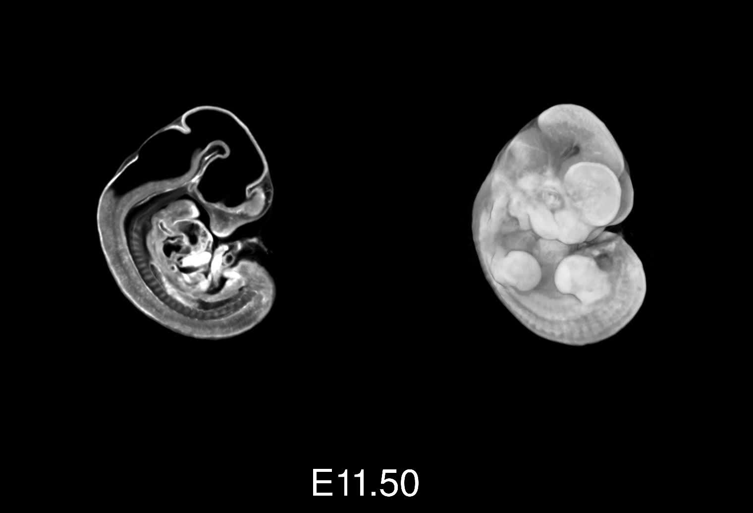
4D atlas of the mouse embryo for precise morphological staging. - Abstract  - Europe PMC
4D atlas of the mouse embryo for precise morphological staging. - Abstract - Europe PMC

The Spatial Mouse Atlas: new insights into cell fate | EMBL

Digital Atlas of Mouse Embryology, CD Atlas

4D atlas of mouse development - YouTube

Mouse Anatomy | FaceBase

4D atlas of the mouse embryo for precise morphological staging |  Development | The Company of Biologists
4D atlas of the mouse embryo for precise morphological staging | Development | The Company of Biologists
eHistology - online annotated histology of the mouse embryo | Biological  Visualisation Community
eHistology - online annotated histology of the mouse embryo | Biological Visualisation Community

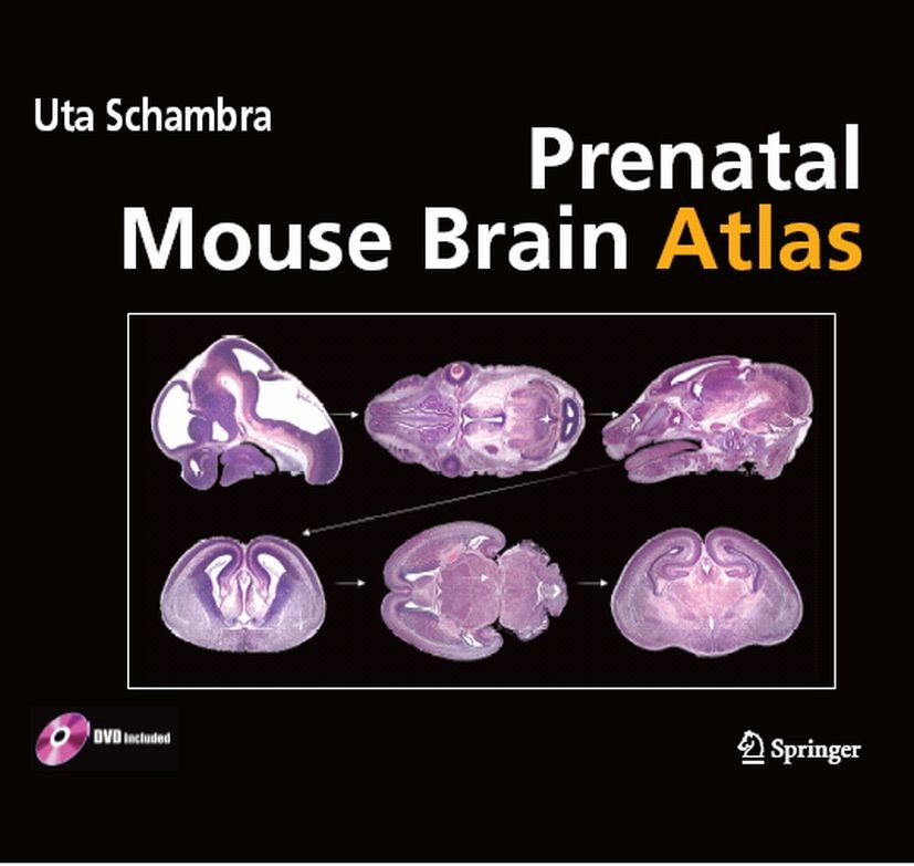
Prenatal Mouse Brain Atlas: Color images and annotated diagrams of:  Gestational Days 12, 14, 16 and 18 Sagittal, coronal and horizontal section  | SpringerLink
Prenatal Mouse Brain Atlas: Color images and annotated diagrams of: Gestational Days 12, 14, 16 and 18 Sagittal, coronal and horizontal section | SpringerLink

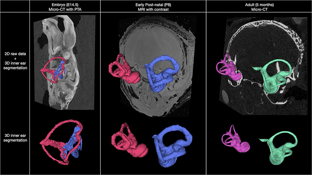
Frontiers | Multimodal Atlas of the Murine Inner Ear: From Embryo to Adult

The venous system of E14.5 mouse embryos—reference data and examples for  diagnosing malformations in embryos with gene deletions - Geyer - 2022 -  Journal of Anatomy - Wiley Online Library
The venous system of E14.5 mouse embryos—reference data and examples for diagnosing malformations in embryos with gene deletions - Geyer - 2022 - Journal of Anatomy - Wiley Online Library

A mouse embryo atlas at E14.5 dpc. This atlas is an average of 16... |  Download Scientific Diagram
A mouse embryo atlas at E14.5 dpc. This atlas is an average of 16... | Download Scientific Diagram

A High-Resolution Anatomical Atlas of the Transcriptome in the Mouse Embryo  | PLOS Biology
A High-Resolution Anatomical Atlas of the Transcriptome in the Mouse Embryo | PLOS Biology

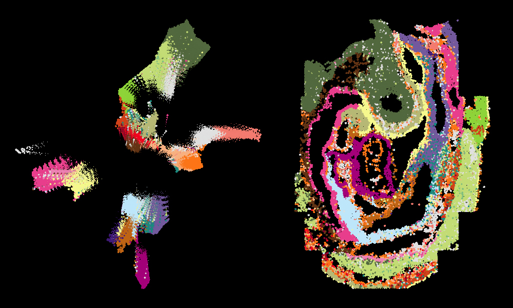
Mouse Organogenesis Spatiotemporal Transcriptomic Atlas - MOSTA -  小鼠胚胎发育时空转录组图谱
Mouse Organogenesis Spatiotemporal Transcriptomic Atlas - MOSTA - 小鼠胚胎发育时空转录组图谱

The Atlas of Mouse Development - 1st Edition